In today’s healthcare environment, intravenous therapy ranks among the most frequently performed medical procedures, with nearly every hospitalized patient requiring venous access at some point during their stay. However, traditional IV needles pose significant safety hazards during use, particularly the risk of needlestick injuries. Statistics show that healthcare workers experience over one million needlestick accidents annually, with a substantial percentage stemming from IV insertion procedures. These accidental injuries not only risk transmission of bloodborne pathogens like hepatitis B, hepatitis C, and HIV, but also create tremendous psychological stress and financial burden for healthcare workers.
The introduction of safety IV catheters has completely transformed this landscape. These innovative medical devices maintain all the functionality of traditional catheters while integrating intelligent safety protection mechanisms. Once venous puncture is complete, the safety device automatically activates, fully encasing or retracting the sharp needle, fundamentally eliminating the possibility of secondary needlestick injuries. This safety IV catheter insertion technique not only protects healthcare workers’ occupational safety but also enhances the patient treatment experience through improved IV catheter safety procedures.
Safety IV catheters represent one of the most significant advances in venous access technology over the past two decades. These sophisticated medical devices have revolutionized intravenous therapy by addressing the critical issue of needlestick injuries that have long plagued healthcare settings. The integration of automatic safety mechanisms with traditional catheter functionality has established a new standard of care that prioritizes both patient outcomes and healthcare worker protection.
The development of safety IV catheters emerged from extensive research into occupational hazards in healthcare environments. Studies consistently demonstrated that conventional IV insertion procedures accounted for a substantial percentage of needlestick injuries among nurses and physicians. These incidents not only posed immediate physical risks but also created significant psychological stress and financial burdens for healthcare institutions through testing, prophylactic treatments, and potential litigation costs.
Understanding Safety IV Catheters
What is a Safety IV Catheter?
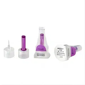
A safety IV catheter is a meticulously engineered medical device that combines the essential functions of traditional IV catheters with advanced safety protection technology. The core innovation lies in its integrated safety mechanism, which automatically protects the needle after venous puncture completion, preventing accidental needlestick injuries from occurring.
Structurally speaking, safety IV catheters contain multiple precision components. The main structure consists of materials with excellent biocompatibility, ensuring optimal compatibility with human tissue. The catheter portion uses soft polyurethane or silicone materials that provide sufficient strength for puncture while maintaining excellent flexibility to minimize vascular wall irritation. The needle component features specialized design that not only maintains sharpness to ensure puncture success rates but also integrates complex safety devices.
The safety mechanism operates based on precision mechanical design. When healthcare providers complete venous puncture and prepare to withdraw the needle, they simply press or push a specific activation button, and the safety device immediately engages. Depending on the design type, the needle either automatically retracts into a protective sheath or becomes completely encased by a sturdy cover. Once the safety mechanism activates, it cannot be reversed, ensuring absolute safety.
A safety IV catheter differs fundamentally from traditional IV catheters in its approach to risk management. While conventional catheters rely primarily on careful handling and disposal procedures to prevent injuries, safety catheters build protection directly into the device itself. This proactive approach represents a paradigm shift from relying on human behavior modification to implementing engineering controls.
The sophisticated engineering behind these devices involves careful consideration of multiple factors including activation force requirements, mechanism reliability, and user feedback systems. Manufacturers conduct extensive testing to ensure that safety mechanisms activate consistently under various conditions while remaining intuitive for healthcare providers to operate. The balance between ease of use and security is crucial, as overly complex mechanisms may discourage proper usage, while overly simple ones might activate prematurely.
Key Components
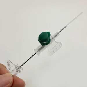
The Catheter Hub serves as the control center of the entire safety IV catheter system. This seemingly simple component actually integrates complex mechanical structures and precision engineering design. The catheter hub must not only house the core components of the safety mechanism but also ensure perfect compatibility with standard medical equipment. Modern catheter hubs are manufactured from engineering plastics with excellent strength and chemical corrosion resistance. Their internal structure includes the safety device trigger mechanism, locking system, and status indicators.
Designers developed the catheter hub with ergonomic principles in mind, ensuring healthcare providers can comfortably and securely grip the device. The surface typically features anti-slip texture design, providing excellent grip even when wearing gloves. Color-coding systems help quickly identify different catheter sizes, improving operational efficiency and safety.
The Flexible Catheter is the critical component that directly contacts the patient’s vascular system, and its material selection and design have direct impact on patient comfort and treatment effectiveness. Modern safety IV catheter tubing is primarily manufactured from polyurethane or silicone biocompatible materials. These materials undergo rigorous biocompatibility testing to ensure they won’t cause adverse reactions during extended placement.
The catheter’s geometric shape has been carefully optimized with a tapered design that facilitates puncture while reducing vascular damage. Wall thickness control is extremely precise, providing sufficient strength to withstand puncture forces while maximizing internal diameter to ensure excellent fluid flow characteristics. The surface undergoes special treatment for good lubricity, reducing insertion resistance and patient discomfort.
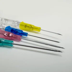
The Safety Needle represents the most critical innovative component of the entire system. Compared to traditional needles, safety needles must not only possess excellent puncture performance but also integrate complex safety protection mechanisms. The needle is manufactured from medical-grade stainless steel with special heat treatment processes ensuring optimal hardness and toughness balance. The needle tip uses ultra-precision grinding technology to form a perfect sharp angle that easily penetrates skin and vascular walls while minimizing tissue damage.
Safety mechanism integration presents the core design challenge for safety needles. Engineers have developed various safety technologies, including passive and active categories. Passive safety mechanisms activate automatically upon needle withdrawal without requiring additional operations, while active mechanisms require healthcare providers to manually trigger activation. Each design has specific application scenarios and advantages.
The Flash Chamber provides intuitive confirmation of successful venous puncture. This transparent small chamber appears simple in design but actually requires precise engineering calculations. The chamber volume must be small enough to ensure rapid blood return while being large enough to provide clearly visible blood confirmation. Material selection uses high-transparency medical-grade plastics ensuring clear blood flow observation under various lighting conditions.
The flash chamber’s geometric shape has also been optimized with special internal surface treatments ensuring blood can quickly enter and remain clearly visible. Some advanced designs include blood return enhancement technology using fine internal surface structures to guide blood flow, improving visibility and speed of blood return.
The Luer Lock Connection ensures safe, reliable connection with various infusion equipment. This standardized connection method uses threaded locking design capable of withstanding considerable pulling and torsional forces. The connection portion uses precision machining technology ensuring perfect compatibility with standard luer fittings. Sealing performance is guaranteed through multiple design features including precise thread matching and dedicated sealing rings.
Modern luer lock designs also consider usability factors, incorporating large-diameter operating rings for easy manipulation even while wearing gloves. Surface texture design provides excellent grip, preventing slippage at critical moments. Some premium products integrate connection status indicators providing color changes or audible feedback to confirm proper connection.
Pre-Insertion Preparation
Equipment Assembly
Equipment assembly represents the foundational phase for ensuring successful safety IV catheter use, requiring a systematic approach and meticulous attention to detail. The quality of preparation work directly influences subsequent operation success rates and patient safety.
Physician Order Verification and Indication Confirmation serves as the starting point and most critical safety check of the entire process. Healthcare providers must carefully review physician-ordered IV therapy orders, including medication types, dosages, administration routes, infusion rates, and other detailed information. This process involves not just simple verification but professional assessment of treatment plan appropriateness.
Patient identification uses a dual identification system, typically including name verification along with medical record number or birth date confirmation. This practice effectively prevents medication errors and treatment plan confusion. Modern hospitals commonly use barcode or RFID technology to assist with identification confirmation, further improving accuracy and efficiency. Healthcare providers must communicate directly with patients, confirming their understanding of upcoming treatment and addressing relevant questions.
Vascular Assessment represents a professional process requiring rich clinical experience. Healthcare providers must comprehensively evaluate patient vascular conditions, including vessel caliber, elasticity, depth positioning, and surrounding tissue status. Ideal puncture sites should feature straight, large, elastic veins while avoiding joint activity areas, scar tissue, and previous puncture sites.
For patients with poor vascular conditions, special vessel visualization techniques may be necessary, such as local warming, gentle massage, or vascular visualization device use. Elderly patients, chemotherapy patients, or long-term hospitalized patients often have fragile vessels requiring particularly careful handling approaches.
Equipment Selection and Preparation requires personalized configuration based on specific patient conditions and treatment needs. IV catheter size selection follows the “minimum effective principle,” meaning selecting the smallest size device that meets treatment requirements. Oversized catheters increase vascular injury risk and patient discomfort, while undersized catheters may affect infusion efficiency.
Common sizes include 14G, 16G, 18G, 20G, 22G, etc., with each size having specific application ranges. 14G-16G are primarily used for emergency and rapid infusion scenarios; 18G-20G suit general inpatient routine treatment; 22G and smaller sizes are mainly used for pediatric patients or elderly patients with poor vascular conditions.
Ancillary Supply Preparation is equally important, including various antiseptic products, securing materials, and emergency equipment. Antiseptic selection typically includes 2% chlorhexidine alcohol solution or 70% isopropyl alcohol, products with broad-spectrum antimicrobial effects and rapid action characteristics. Sterile gauze, medical tape, transparent dressings, and other securing materials should be selected based on patient skin conditions and expected dwelling time.
Patient preparation extends beyond physical aspects to include psychological readiness. Many patients experience anxiety about IV insertion procedures, particularly those with previous negative experiences. Healthcare providers should take time to explain the procedure, address concerns, and provide emotional support. This communication not only improves patient cooperation but also contributes to better overall outcomes.
The preparation phase also involves environmental considerations. The procedure area should be well-lit, clean, and equipped with all necessary supplies within easy reach. Adequate lighting is crucial for successful vein identification and puncture, while maintaining a sterile environment prevents contamination and reduces infection risk.
Site Selection and Preparation
The Art and Science of Vessel Selection combines anatomical knowledge, clinical experience, and individual patient characteristics in comprehensive consideration. The upper extremity venous system provides multiple selectable puncture sites for IV therapy, with each location offering unique advantages and limitations.
The Dorsal Hand Venous Network typically represents the preferred puncture site, particularly for patients requiring short-term infusion therapy. This region’s veins are relatively superficial, easily identified and punctured, with minimal impact on patient daily activities. Dorsal hand veins include major branches like the cephalic vein, basilic vein, and dorsal venous arch, vessels that are usually straight with good stability. However, dorsal hand skin is relatively thin, potentially unsuitable for patients requiring irritating medication infusions.
Forearm Veins provide ideal choices for medium to long-term IV therapy. The cephalic and basilic veins in the forearm segment are usually large with sufficient blood flow, capable of accommodating various types of infusion therapy. This region is distant from joints, making it less likely for patient movement to pull the catheter, supporting puncture site stability. Forearm vein selection also requires consideration of patient occupational characteristics and lifestyle habits; for patients requiring frequent bilateral hand use for work, the non-dominant side should be prioritized.
Antecubital Veins, while large and prominent, typically aren’t first-choice sites for indwelling catheters. This region’s veins are mainly used for short-term rapid infusions or blood collection procedures. Due to location in joint activity areas, long-term placement easily causes catheter displacement or kinking, increasing complication risks. Antecubital veins should only be considered when other sites are unsuitable, requiring special attention to securing methods and activity restrictions.
Special Population Vascular Selection Strategies require individualized approaches. Pediatric patients have small vessels with more subcutaneous fat, making vein identification difficult; typically, superficial veins of the dorsal hand or foot are selected. Elderly patients have decreased vascular elasticity and tendency to roll, requiring more careful operative techniques. Obese patients often have deeper veins, potentially requiring palpation techniques or vascular visualization devices for location assistance.
Standardized Skin Preparation Protocol represents a key element in infection prevention that must be strictly executed according to aseptic technique principles. Preparation begins with selecting appropriate antiseptics; currently, 2% chlorhexidine alcohol solution is most commonly used clinically, offering rapid antimicrobial action, sustained bacterial inhibition, and minimal skin irritation characteristics.
The antiseptic area should extend at least 5 centimeters in diameter around the puncture point. The antiseptic method uses spiral application, gradually expanding from center to periphery, avoiding application from contaminated to clean areas. The antiseptic process requires 2-3 repetitions, using new sterile cotton balls or gauze each time to ensure adequate antimicrobial effectiveness.
The Importance of Drying Time is often overlooked but extremely critical. Antiseptics require adequate contact time to achieve optimal antimicrobial effects, typically requiring 30-60 seconds of natural evaporation time. Premature puncture not only affects antiseptic effectiveness but residual antiseptic may cause chemical irritation to vessels and tissues. During the drying wait period, healthcare providers can conduct final equipment checks and mental preparation.
Site preparation also involves patient positioning to optimize vein access and comfort. The selected extremity should be positioned at or slightly below heart level to promote venous filling. Adequate support should be provided to prevent patient fatigue and involuntary movement during the procedure. Pillows or positioning devices can significantly improve both patient comfort and procedural success rates.
Environmental factors such as room temperature can significantly affect vein visibility and accessibility. Cool temperatures cause vasoconstriction, making veins less prominent and more difficult to puncture. Warming the extremity with warm towels or heating pads for several minutes before attempted insertion can improve success rates, particularly in patients with poor peripheral circulation.
Step-by-Step Insertion Technique
The Insertion Process
Catheter Positioning and Angle Control represents the technical core of successful puncture, requiring precise hand-eye coordination and rich clinical experience. Correct puncture angle is crucial for successful single-attempt puncture; overly steep angles easily penetrate the posterior vessel wall, while overly shallow angles may fail to enter the vessel lumen.
Initial Puncture Angle should be controlled between 15-30 degrees, with specific angle selection depending on vessel depth and caliber. Superficial vessels require smaller puncture angles, typically between 15-20 degrees; deeper vessels may require slightly larger angles but rarely exceed 30 degrees. Puncture angle determination requires combining palpation sensation with visual observation; experienced healthcare providers can determine optimal puncture angles by feeling vessel pulsation and elasticity.
The Essence of Puncture Technique lies in stability, confidence, and continuity. Puncture movement should be performed in one smooth motion, avoiding hesitation and repeated adjustments. The needle bevel must face upward, reducing damage to vascular intima and improving puncture success rates. Puncture speed should be moderate; excessive speed risks loss of control, while insufficient speed increases patient suffering and vascular spasm risk.
The Moment of Skin Penetration requires overcoming epidermal resistance, creating a distinct “pop” sensation. Experienced operators can accurately perceive this moment and immediately adjust puncture direction toward the vessel. After skin penetration, the needle should continue advancing forward, seeking resistance sensation from the vessel wall. When the needle tip contacts the vessel wall, there will be slight resistance increase, marking the critical moment for vessel puncture.
Vessel Wall Penetration Technique requires delicate tactile perception and precise force control. Penetrating the vessel wall requires certain pushing force but must be controlled within appropriate ranges to avoid excessive force causing contralateral wall injury. Successful vessel wall penetration is marked by sudden resistance disappearance while simultaneously observing blood return in the flash chamber.
The Importance of Blood Return Confirmation serves not only as a puncture success indicator but also as important reference for subsequent operations. Blood return speed and color can reflect vessel condition and puncture accuracy. Arterial blood appears bright red with pulsatile flow; venous blood appears dark red with continuous flow. If blood return is very slow or color is abnormal, puncture position and depth require reassessment.
The Critical Step of Catheter Advancement represents the most skill-requiring phase of the entire operation. After observing blood return, the entire assembly (needle and catheter together) should first advance 1-2 millimeters forward, ensuring the catheter tip also enters the vessel lumen. This step, called “floating technique,” is key to preventing catheter obstruction or damage during advancement.
Catheter Threading Technique requires extremely gentle and smooth movements. Hold the needle steady while using the other hand to gently thread the catheter along the needle into the vessel. If resistance is encountered during advancement, never force progression; instead, assess the cause of resistance. Possible causes include catheter tip meeting vessel wall, vascular spasm, or catheter kinking.
When advancing the catheter, maintain direction parallel to the vessel axis, avoiding lateral force application. Advancement speed should be uniform and slow, allowing the vessel time to adapt. If blood return stops during advancement, this may indicate improper catheter position or vascular spasm, requiring immediate cessation of advancement and reassessment.
The insertion process requires maintaining sterile technique throughout the entire procedure. Any contamination of the catheter or insertion site can lead to serious infections. Healthcare providers must remain vigilant about maintaining the sterile field and avoiding contact between sterile components and non-sterile surfaces.
Patient communication during insertion is equally important. Providing reassurance, explaining sensations the patient might experience, and maintaining a calm demeanor can significantly reduce patient anxiety and improve cooperation. Some patients may experience vasovagal responses during insertion, so healthcare providers should be prepared to recognize and manage these reactions promptly.
Safety Mechanism Activation
Safety Mechanism Activation Timing represents a key element in ensuring operational safety and must occur immediately after complete catheter placement. Delayed safety mechanism activation increases accidental needlestick injury risk, while premature activation may affect proper catheter placement. Ideal activation timing occurs after confirming correct catheter position and unobstructed blood flow, at the moment of preparing needle withdrawal.
Operating Methods for Different Safety Mechanism Types each have unique characteristics requiring healthcare provider familiarity with specific requirements of various products. Passive Safety Mechanisms feature the most convenient design, automatically activating when the needle withdraws to a specific position. This design’s advantage lies in requiring no additional operational steps, reducing human error possibilities. During operation, healthcare providers simply withdraw the needle normally; when the needle tip completely exits the catheter, the safety device automatically encases the needle.
Active Safety Mechanisms require healthcare providers to manually trigger activation. Common activation methods include pressing activation buttons, pushing slides, or rotating safety covers. This design’s advantage lies in providing operators complete control, allowing selection of optimal activation timing. Operation requires using one hand to stabilize the catheter while the other hand operates the safety device, ensuring coordinated movement.
The Importance of Activation Confirmation cannot be overlooked and must be confirmed through both visual and auditory means that the safety mechanism has properly engaged. After correct activation, the needle should be completely encased or retracted by the safety device, unable to be re-exposed. Many modern products feature activation confirmation indicators such as color changes, audible feedback, or position locking.
Response Measures for Activation Failure require advance preparation and rapid response. If the safety mechanism cannot activate normally, never force operation, as this may damage equipment or cause accidental injury. The correct approach is to immediately treat the entire device as a traditional sharp, following standard sharps disposal procedures for safe handling.
Simultaneously prepare backup safety catheters and restart puncture procedures. For devices with activation failure, report according to healthcare institution adverse event reporting procedures and notify equipment suppliers and regulatory agencies. This information feedback holds important significance for product quality improvement and industry safety standard enhancement.
Post-Activation Safety Mechanism Handling requires special attention to maintaining sterile environment and equipment integrity. Activated safety devices should not be disassembled or modified and must remain in original condition until final disposal. Even with activated safety mechanisms, exercise caution when handling equipment, avoiding unnecessary contact and collision.
The psychological impact of successful safety mechanism activation should not be underestimated. Healthcare workers report significantly reduced anxiety and stress levels when using safety devices compared to conventional catheters. This psychological benefit translates into improved job satisfaction and potentially better patient care outcomes.
Training programs must emphasize the importance of never attempting to defeat or bypass safety mechanisms. Even when devices appear to malfunction, healthcare providers should resist the temptation to force activation or modify the device. Such actions can lead to serious injuries and defeat the entire purpose of using safety equipment.
Post-Insertion Care
Immediate Assessment
Catheter Position Verification represents the primary step in ensuring treatment safety and effectiveness. Successful venous puncture requires not only catheter entry into the vessel lumen but also ensuring correct, stable catheter position with good function. The verification process includes multiple assessment aspects, each with specific significance and methods.
Blood Return Testing provides the most direct position confirmation method. Gently aspirate with a syringe or infusion device; you should easily draw bright red venous blood. Blood return speed, color, and volume all provide important diagnostic information. Rapid, abundant dark red blood return typically indicates correct and patent catheter position. If blood return is slow, minimal, or abnormally colored, this may suggest improper catheter position, vascular spasm, or other issues.
Saline Flush Testing further verifies catheter patency and correct position. Using a 10ml syringe, slowly inject a small amount of normal saline; normally there should be no resistance, and patients should not experience significant discomfort. During flushing, carefully observe the puncture site ensuring no fluid leakage or swelling. If injection resistance significantly increases or the puncture site develops swelling, this suggests possible improper catheter position or extravasation.
Early Identification of Infiltration and Extravasation is crucial for preventing IV catheter complications. Infiltration refers to infusion entering tissue spaces surrounding vessels, usually caused by improper catheter position or vessel wall damage. Extravasation specifically refers to irritating or corrosive medications entering extravascular tissue, potentially causing serious tissue necrosis.
Early identification keys lie in systematic observation and assessment. First, check the puncture site for swelling, redness, warmth, or pain indicating inflammation. Gently palpate around the puncture site, feeling for induration or edema. Inquire about patient sensations, including presence of pain, numbness, tingling, or other abnormal feelings.
Vascular Access Function Assessment includes infusion rate and pressure testing. Normal IV catheters should support required infusion rates without significant resistance or pressure increases. If higher infusion pressures are needed to maintain normal rates, this may suggest partial catheter obstruction or improper positioning.
Patient Comfort Assessment is equally important and often directly relates to catheter placement success. Properly placed catheters should cause minimal patient discomfort once initial insertion pain subsides. If patients report persistent pain, pressure sensation, or movement restrictions, this may indicate catheter malposition or vascular complications.
Documentation and Communication form essential components of post-insertion care. Accurate documentation should include insertion time, catheter size and type, insertion site location, number of attempts required, patient response, and any complications encountered. This information not only supports continuity of care but also provides valuable data for quality improvement initiatives.
Communication with patients and family members should include explanation of care requirements, signs and symptoms to report, and activity restrictions if any. Patient education empowers individuals to participate in their care and helps ensure early detection of potential complications.
Securing and Maintenance
Catheter Securement Techniques play a crucial role in preventing complications and ensuring treatment success. Proper securement prevents catheter migration, reduces infection risk, and improves patient comfort during the dwelling period. Modern securement approaches have evolved beyond traditional tape methods to include specialized securement devices designed specifically for IV catheters.
Transparent Dressing Application provides several advantages including moisture barrier protection, visual inspection capability, and secure adhesion. The dressing should completely cover the insertion site while allowing clear visualization of the catheter hub and surrounding tissue. Application technique requires careful attention to avoid air bubbles or wrinkles that could compromise adhesion or create bacterial harboring sites.
When applying transparent dressings, ensure the insertion site is completely dry and free from residual antiseptic. Center the dressing over the insertion site and apply with smooth, even pressure from center outward to eliminate air pockets. The dressing should extend beyond the catheter hub to provide adequate stabilization while not interfering with IV tubing connections.
Catheter Hub Stabilization prevents movement that could lead to mechanical complications or patient discomfort. The hub should be secured in a position that maintains straight alignment with the vessel and minimizes tension on the catheter. Various commercial securement devices are available that provide superior stabilization compared to traditional tape methods.
These specialized devices distribute securement forces over larger surface areas, reducing pressure points and skin breakdown risk. They also typically allow for easy access to the catheter hub for flushing or medication administration while maintaining secure positioning throughout the dwelling period.
IV Tubing Management requires attention to prevent inadvertent catheter displacement or patient entanglement. Tubing should be secured with sufficient slack to allow normal patient movement while preventing excessive tension on the catheter. Loop formations in the tubing can provide strain relief and reduce direct pulling forces on the insertion site.
Consider patient positioning and mobility needs when planning tubing routes. For ambulatory patients, tubing should be secured to prevent catching on obstacles or dragging. For bedridden patients, ensure tubing doesn’t become trapped under the patient or caught in bed mechanisms.
Site Inspection Protocols should be established and followed consistently throughout the catheter dwelling period. Regular assessment allows for early detection of complications and prompt intervention when needed. Inspection frequency typically ranges from every 4-8 hours depending on patient acuity and institutional protocols.
During each inspection, assess for signs of infiltration, extravasation, phlebitis, or infection. Document findings consistently using standardized assessment tools when available. Any abnormal findings should prompt immediate evaluation and appropriate interventions.
Patient Activity Guidelines should balance treatment requirements with quality of life considerations. Most patients with properly secured IV catheters can engage in normal activities with minimal restrictions. However, certain precautions may be necessary depending on catheter location and treatment requirements.
Educate patients about protecting the IV site during activities of daily living, including bathing, dressing, and sleeping. Provide guidance on recognizing signs that require immediate attention and when to seek help from healthcare providers.
Safety Protocols and Best Practices
Infection Prevention Strategies
Comprehensive Aseptic Technique forms the foundation of infection prevention in IV catheter procedures. This approach goes beyond simple hand hygiene and sterile glove use to encompass a complete system of contamination prevention measures. Every aspect of the procedure, from equipment preparation through catheter removal, must adhere to strict aseptic principles.
Hand Hygiene Excellence represents the single most important infection prevention measure. Healthcare providers must perform thorough hand hygiene immediately before and after every patient contact, using either alcohol-based hand rub or soap and water depending on the clinical situation. Hand hygiene should also be performed before donning gloves and after glove removal, as gloves can develop microscopic perforations during use.
The technique of hand hygiene is as important as frequency. Alcohol-based preparations should be applied to cover all hand surfaces and rubbed vigorously until completely dry, typically requiring 20-30 seconds. When using soap and water, hands should be wetted first, soap applied, and all surfaces scrubbed for at least 15 seconds before thorough rinsing and drying with clean towels.
Sterile Field Maintenance requires constant vigilance throughout the procedure. Once established, the sterile field must be monitored continuously to ensure no contamination occurs. This includes avoiding contact between sterile items and non-sterile surfaces, preventing sterile items from falling below waist level, and ensuring sterile packages are opened properly to maintain contents sterility.
Healthcare providers must understand the principles of sterile technique including concepts of sterile versus clean, contamination recognition, and appropriate responses to potential contamination events. When contamination occurs or is suspected, the affected items must be considered non-sterile and replaced before continuing the procedure.
Catheter Site Care Protocols extend infection prevention beyond the insertion procedure to include ongoing maintenance throughout the dwelling period. Daily site assessment should include inspection for signs of local infection such as redness, swelling, warmth, tenderness, or purulent drainage. Documentation of these assessments provides important data for trending and early intervention.
Dressing Change Procedures should follow standardized protocols designed to minimize contamination risk while ensuring adequate site protection. Dressing changes are typically required when dressings become loose, wet, soiled, or compromised in any way. Some institutional protocols also mandate routine dressing changes at specified intervals regardless of dressing condition.
During dressing changes, strict aseptic technique must be maintained. Old dressings should be removed carefully to avoid catheter displacement, the site should be cleaned with appropriate antiseptic if needed, and new dressings applied using sterile technique. The procedure should be performed by healthcare providers with appropriate training and competency validation.
Catheter Flushing and Medication Administration present ongoing infection risks that require careful attention to aseptic technique. All hub connections should be disinfected with appropriate antiseptic before access, using friction and adequate contact time to ensure effectiveness. Needleless connectors should be scrubbed with 70% alcohol for at least 15 seconds and allowed to dry before access.
Environmental Considerations play important roles in infection prevention. The procedure environment should be clean, well-lit, and free from unnecessary traffic. Equipment and supplies should be stored properly to maintain cleanliness and prevent contamination. Some procedures may benefit from being performed in designated clean procedure areas rather than at the bedside.
Complication Prevention and Management
Infiltration and Extravasation Prevention requires proactive monitoring and risk factor assessment throughout the catheter dwelling period. Risk factors for these complications include catheter size relative to vessel caliber, insertion technique quality, patient activity level, and types of medications being administered.
Early Recognition Systems should be implemented to identify infiltration and extravasation at the earliest possible stages. This includes patient education about symptoms to report, regular nursing assessments using standardized scales, and appropriate response protocols when complications are detected. Early intervention significantly reduces the severity of tissue damage and improves patient outcomes.
Assessment scales provide objective measures for evaluating infiltration severity and guiding intervention decisions. These tools typically consider factors such as tissue edema extent, skin color changes, temperature variations, and patient pain levels. Consistent use of standardized scales improves communication between healthcare providers and ensures appropriate escalation of care.
Phlebitis Prevention focuses on minimizing vessel irritation through appropriate catheter selection, proper insertion technique, and careful medication administration practices. Risk factors include catheter dwelling time, insertion technique, catheter material and size, and characteristics of infused solutions.
Catheter Rotation Policies help prevent phlebitis by limiting vessel exposure time and allowing healing of previously accessed sites. Most guidelines recommend catheter replacement every 72-96 hours for peripheral IVs in adult patients, with modifications based on individual patient factors and catheter condition assessments.
pH and Osmolarity Management of infused solutions can significantly impact phlebitis development. Highly acidic or alkaline solutions, as well as those with extreme osmolarity, increase vessel irritation risk. When such solutions must be administered through peripheral IVs, consider dilution when possible or alternative access routes for problematic medications.
Air Embolism Prevention requires attention to proper priming of all IV tubing and careful monitoring of infusion systems. All air should be removed from tubing before connection to patients, and infusion pumps should be equipped with air detection systems when available. Patient positioning during line changes and careful attention to connection security help minimize air entry risk.
Infection Prevention Beyond Site Care includes attention to IV fluid preparation, tubing changes, and hub access procedures. IV fluids should be prepared using aseptic technique in appropriate environments, with attention to beyond-use dating and storage requirements. Tubing changes should follow manufacturer recommendations and institutional protocols to balance infection risk with cost considerations.
Troubleshooting Common Issues
Managing Failed Insertion Attempts
First Attempt Analysis should occur immediately when initial insertion fails, focusing on identifying correctable factors that contributed to the failure. Common causes include inappropriate site selection, suboptimal insertion angle, inadequate vessel stabilization, or patient movement during the procedure. Understanding failure mechanisms guides decisions about repeat attempts and technique modifications.
Site Reassessment may reveal better alternatives that were not initially apparent. Patient positioning changes, improved lighting, or warming techniques might make previously unsuitable vessels more accessible. Consider moving to different anatomical locations rather than repeated attempts at the same site, which can cause vessel damage and reduce success probability for subsequent tries.
Technique Modification based on initial attempt experience often improves success rates. This might include angle adjustments, approach direction changes, or stabilization technique improvements. However, avoid making multiple complex changes simultaneously, as this makes it difficult to identify which modifications are beneficial.
Equipment Considerations should include evaluation of catheter size appropriateness for the selected vessel. Smaller gauge catheters may be necessary for difficult access situations, even if larger sizes would be preferred for clinical reasons. The clinical team should weigh the benefits of immediate access establishment against optimal flow rate requirements.
Alternative Access Strategies should be considered when peripheral access proves challenging. This might include ultrasound-guided peripheral access, external jugular access, or central venous access depending on clinical requirements and provider capabilities. Early recognition of difficult access situations and appropriate escalation prevents multiple failed attempts and patient discomfort.
Patient Comfort Management during multiple attempt situations requires special attention. Adequate pain management, emotional support, and clear communication about the situation help maintain patient cooperation and reduce anxiety. Consider involving additional skilled personnel or implementing comfort measures before continuing with access attempts.
Device Malfunction Management
Safety Mechanism Failure represents a serious concern requiring immediate appropriate response. Never attempt to force activation of malfunctioning safety devices, as this may cause device damage or create injury hazards. Instead, treat the device as a conventional sharp and follow institutional sharps handling procedures.
Immediate Actions should include securing the malfunctioning device to prevent injury, notifying appropriate personnel, and preparing alternative equipment for patient care continuation. Document the malfunction thoroughly including device lot numbers, expiration dates, and specific failure characteristics observed.
Reporting Requirements typically include internal incident reporting through institutional quality assurance systems and external reporting to device manufacturers and regulatory agencies when appropriate. These reports contribute to device improvement efforts and may identify patterns requiring broader interventions.
Product Recall Response procedures should be established in advance to ensure rapid, appropriate responses when manufacturers issue safety alerts or recalls. This includes inventory identification systems, user notification procedures, and alternative product preparation.
Catheter Occlusion Management requires systematic assessment to determine occlusion type and appropriate interventions. Mechanical occlusions might respond to position changes or gentle flushing, while thrombotic occlusions may require specific dissolution agents. Always follow institutional protocols for occlusion management and avoid excessive pressure that could cause catheter rupture.
Hub Connection Problems can usually be resolved through careful cleaning and inspection of connection surfaces. Debris, dried blood, or medication residue can interfere with proper connections. Clean connections with appropriate antiseptic and ensure proper alignment before attempting reconnection. If threading problems persist, inspect for cross-threading or damaged threads that might require equipment replacement.
Flow Rate Issues may result from various causes including catheter kinking, patient positioning, infiltration, or pump malfunctions. Systematic troubleshooting should begin with simple interventions like position changes and progress through more complex assessments. Never force fluids through resistant catheters, as this may cause catheter rupture or tissue damage.
Quality Improvement and Training
Competency Development Programs
Foundational Knowledge Requirements for healthcare providers using safety IV catheters extend beyond basic insertion techniques to include comprehensive understanding of vascular anatomy, infection control principles, complication recognition, and device-specific operating procedures. Educational programs should address both theoretical knowledge and practical skill development through structured learning experiences.
Anatomical Understanding forms the basis for successful catheter insertion and includes detailed knowledge of upper extremity venous anatomy, vessel characteristics at different ages and health states, and factors affecting vessel accessibility. Providers should understand anatomical variations that might affect insertion success and be able to identify optimal puncture sites based on individual patient characteristics.
Pathophysiology Comprehension helps providers understand complication development mechanisms and appropriate prevention strategies. This includes understanding of inflammatory responses, coagulation processes, fluid dynamics, and tissue healing mechanisms. Such knowledge enables providers to make informed decisions about catheter selection, insertion techniques, and ongoing care strategies.
Technical Skill Development requires hands-on practice with various catheter types and safety mechanisms. Training programs should provide opportunities to practice with different devices under supervised conditions, allowing learners to develop muscle memory and confidence with equipment operation. Simulation-based training can provide safe learning environments for skill development without patient risk.
Competency Assessment Methods should include both knowledge testing and practical skill evaluation. Written examinations can assess theoretical understanding, while practical evaluations should include actual catheter insertion procedures observed by qualified assessors. Assessment should be repeated periodically to ensure skill maintenance and identify learning needs.
Ongoing Education Requirements reflect the evolving nature of IV catheter technology and evidence-based practice recommendations. Healthcare providers should participate in regular updates about new devices, technique improvements, and research findings that might affect clinical practice. Professional development opportunities should be accessible and relevant to current practice needs.
Simulation Training Benefits include risk-free skill practice, exposure to various clinical scenarios, and opportunity for immediate feedback. High-fidelity simulators can reproduce realistic patient conditions including difficult access situations, allowing providers to develop problem-solving skills without compromising patient safety.
Performance Monitoring Systems
Key Performance Indicators for safety IV catheter programs should include metrics that reflect both safety outcomes and clinical effectiveness. Success rate measurements provide important data about technique effectiveness and training program adequacy. These metrics should be tracked consistently and analyzed for trends that might indicate areas needing improvement.
First-Attempt Success Rates represent important quality indicators that reflect provider skill levels, equipment appropriateness, and patient selection factors. High first-attempt success rates indicate effective training programs and appropriate clinical practices, while declining rates may signal need for additional education or technique review.
Needlestick Injury Surveillance provides direct measures of safety device effectiveness and proper usage compliance. Tracking injury rates, circumstances surrounding injuries, and device types involved helps identify areas for improvement and demonstrates program effectiveness. Zero injury goals should be established with supporting systems to achieve and maintain this target.
Catheter-Related Complications tracking helps identify patterns that might indicate system problems or opportunities for improvement. Complication rates should be analyzed by provider, unit, patient population, and device type to identify specific areas needing attention. Trending analysis helps distinguish normal variation from significant changes requiring intervention.
Patient Satisfaction Measures reflect the patient experience with IV catheter procedures and can provide insights into areas for improvement. Patient feedback about pain levels, communication quality, and overall procedure experience helps guide training focus and quality improvement initiatives.
Cost-Effectiveness Analysis should consider not only direct device costs but also indirect costs associated with complications, injuries, and staff time. While safety devices may have higher upfront costs, total cost analysis often demonstrates favorable cost-benefit ratios when all factors are considered.
Benchmark Comparisons with other institutions or published standards help organizations understand their performance relative to industry norms. Participation in quality improvement networks provides access to comparative data and best practice sharing opportunities.
Advanced Considerations
Special Population Management
Pediatric Considerations require significant modifications to standard adult techniques due to anatomical, physiological, and psychological differences. Pediatric vessels are smaller, more fragile, and often more difficult to visualize than adult vessels. Specialized pediatric safety catheters are available in smaller sizes with modified safety mechanisms appropriate for smaller hands and different insertion techniques.
Insertion techniques for pediatric patients often require different approaches including transillumination devices, ultrasound guidance, or specialized positioning techniques. Pain management becomes particularly important, with topical anesthetics, distraction techniques, and family involvement playing crucial roles in successful procedures.
Geriatric Patient Adaptations address age-related changes in vascular structure and function. Elderly patients often have fragile, tortuous vessels with reduced elasticity and tendency to roll during puncture attempts. Skin changes including thinning and reduced subcutaneous tissue require modifications in insertion technique and securement methods.
Anticoagulation considerations are particularly important in elderly populations who commonly receive anticoagulant medications. These patients may have prolonged bleeding times and increased bruising risk, requiring modified techniques and enhanced monitoring protocols.
Oncology Patient Considerations include recognition of treatment-related vascular changes, increased infection risk, and special medication requirements. Chemotherapy patients often have compromised vessels, increased bleeding risk, and may require specialized access devices for vesicant medication administration.
Critical Care Applications may require larger bore catheters for rapid fluid resuscitation or blood product administration. Emergency insertion situations require rapid decision-making while maintaining safety principles. Specialized training for critical care applications helps ensure providers can balance speed requirements with safety considerations.
Technology Integration
Electronic Documentation Systems can improve consistency and completeness of IV catheter-related documentation. Integration with electronic health records allows for standardized assessment forms, automated reminders for catheter assessment, and trending analysis of complications and outcomes.
Barcode Technology helps ensure proper device identification and traceability. Scanning systems can verify correct catheter selection, track insertion dates for rotation scheduling, and provide data for quality improvement initiatives.
Ultrasound-Guided Insertion represents an advancing technology for difficult access situations. Training programs should include ultrasound guidance techniques for providers likely to encounter challenging access situations. Proper technique training and competency validation are essential for successful implementation.
Infusion Pump Integration with smart technology helps prevent medication errors and provides additional safety layers. Understanding pump capabilities and limitations helps providers maximize technology benefits while maintaining clinical judgment about appropriate interventions.
Conclusion
The successful implementation of safety IV catheter programs requires comprehensive attention to multiple factors including device selection, provider training, quality monitoring, and continuous improvement initiatives. Healthcare organizations must commit to creating cultures that prioritize both patient safety and healthcare worker protection while maintaining high standards of clinical care.
Evidence-Based Practice Integration should guide all aspects of safety IV catheter programs from device selection through outcome evaluation. Regular review of current literature helps ensure practices remain current with evolving evidence and technology developments. Professional organizations provide valuable resources for staying current with best practice recommendations.
Multidisciplinary Collaboration enhances program effectiveness by incorporating perspectives from nursing, medicine, risk management, infection control, and quality improvement specialists. Each discipline brings unique expertise that contributes to comprehensive program development and implementation.
Patient-Centered Care remains the ultimate goal of all safety IV catheter initiatives. While healthcare worker safety is crucial, patient outcomes must remain the primary focus of all clinical activities. The best safety programs achieve both goals simultaneously through well-designed systems and properly trained personnel.
Future Developments in safety IV catheter technology continue to evolve, with innovations in materials science, safety mechanism design, and integration with other medical technologies. Healthcare providers should remain open to new developments while maintaining critical evaluation skills to assess new technologies appropriately.
The investment in comprehensive safety IV catheter programs represents both ethical obligation and practical necessity in modern healthcare environments. When properly implemented, these programs protect healthcare workers from occupational injuries while improving patient care quality and organizational risk profiles. Success requires ongoing commitment from leadership, adequate resource allocation, and continuous attention to program effectiveness and improvement opportunities.
Healthcare providers at all levels play crucial roles in program success through their daily commitment to following established procedures, participating in training activities, and contributing to quality improvement initiatives. The collective efforts of all team members create safer healthcare environments that benefit patients, providers, and healthcare organizations alike.
This comprehensive guide should be used in conjunction with manufacturer-specific instructions, institutional policies, and current evidence-based practice guidelines. Healthcare providers should maintain current knowledge through continuing education and professional development activities. Always consult facility policies and current literature for the most up-to-date recommendations and requirements.